
Reading path: Marine organisms and body plans →
A new science guide at a historical Coldwater address. About the earlier site →
Photographic reference
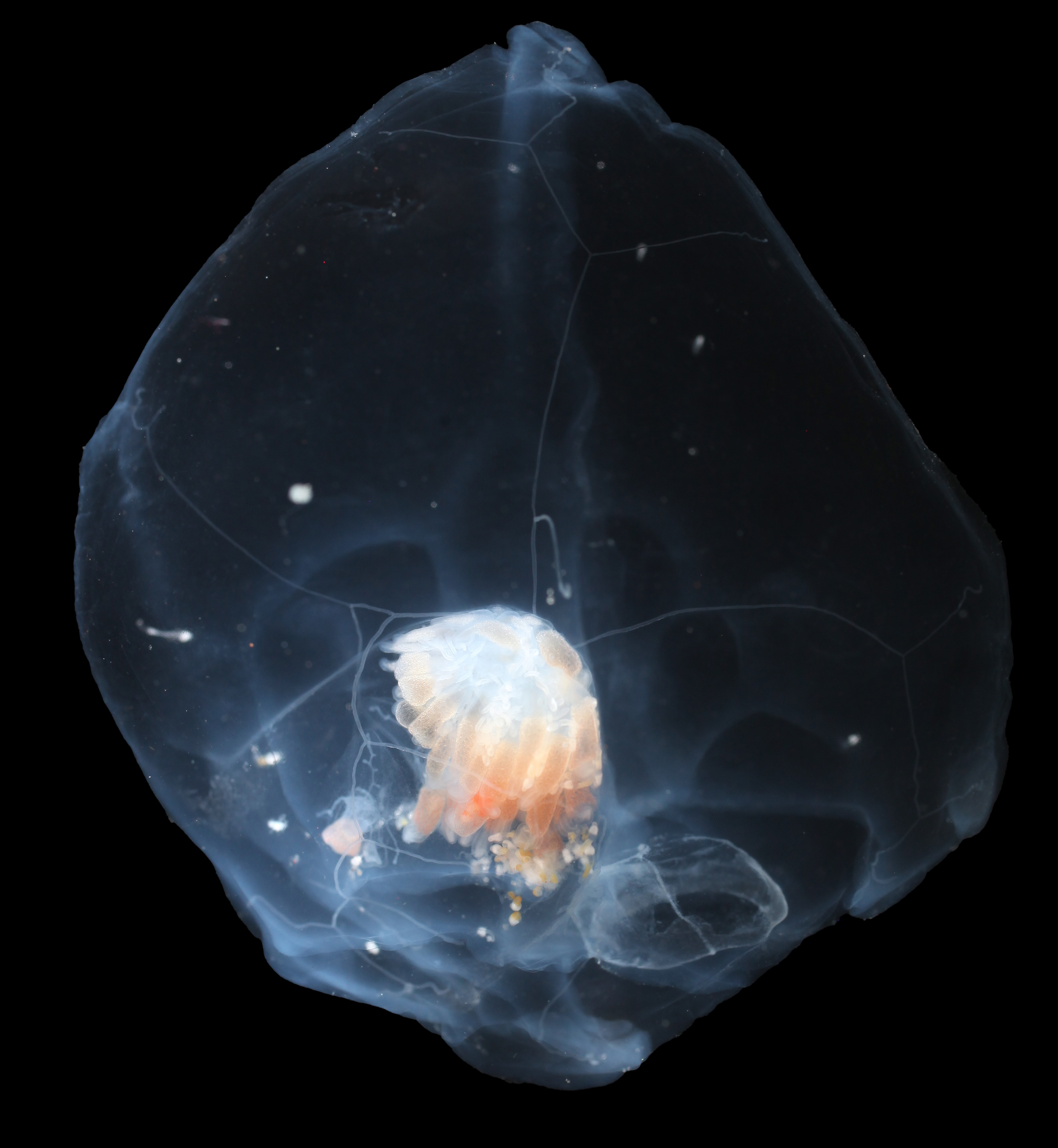
Credit: Allen Collins, NOAA Fisheries National Systematic Laboratory and Smithsonian National Museum of Natural History. Original source · NOAA educational image reuse. None; original photograph displayed proportionally.
What to look for
| Feature | Observation | Comparison or limit |
|---|---|---|
| Organization | Connected specialized zooids share a colony. | They are not independent animals merely traveling together. |
| Feeding | Extended stinging tentacles intercept prey. | Colony and tentacle arrangements vary among groups. |
| Image limits | A frame may show only part of an extended colony. | An unscaled partial frame cannot establish total length. |
Many parts, one connected colony
Siphonophores are colonial hydrozoans made of specialized zooids. The zooids are genetically identical and arise from one fertilized egg, but different zooids perform roles such as feeding, reproduction, or movement. They remain physically integrated; they are not independent animals that happen to travel side by side. [2]
A useful hypothetical comparison is a set of connected modules in which one part captures food while another contributes to movement. The analogy helps explain division of labor, but a siphonophore is a living colony, not a mechanical device. Its coordinated form comes from connected zooids with specialized anatomy.
A Rosacea colony in the Sargasso Sea
A Rosacea siphonophore colony photographed in the Sargasso Sea in April 2006 looked like a single jellyfish at first glance. Its long tentacles extended about a meter from the main body and contracted when disturbed by potential food items. Beadlike structures along the tentacles were identified as nematocysts, stinging cells that immobilize prey. [1]
These are observations from a particular colony, not measurements that define every siphonophore. The example shows how a colony can extend its feeding structures well beyond its central body. A photograph can reveal that spatial arrangement, though it may not capture the full tentacle length or show the behavior that caused a contraction.
Stinging cells and feeding reach
Nematocysts are characteristic of cnidarians, including hydrozoans. When a prey item contacts a tentacle, stinging cells can help immobilize it. In a colony, extended tentacles increase the area that can encounter drifting prey. The Rosacea image records tentacles with beadlike nematocysts; the exact sequence of prey capture is a biological mechanism inferred from the structures and their known function. [1]
This differs from comb-jelly capture, where some species use sticky colloblast cells rather than cnidarian stinging cells. Both groups may look gelatinous in water, so a transparent body or trailing filaments are not enough to tell the mechanism. The distinction depends on anatomy, not simply on a dramatic outline. [2]
Colonies are not all alike
Siphonophore colonies vary in structure and appearance. Some hydrozoans have small medusae or attached hydroid forms instead of a large pelagic colony; siphonophores are one branch within Hydrozoa, not a synonym for the entire class. Colony length and arrangement also vary, so one photographed form should not stand for all. [2]
A photograph may show a chainlike body, a cluster of zooids, or long feeding tentacles. It cannot necessarily establish whether a colony has a gas float, how its zooids divide tasks, or its total length if parts extend beyond the frame. Careful descriptions separate visible features from biological interpretation: connected repeated structures support a colonial reading, while finer conclusions require more anatomical evidence. [1]
Sources & further reading
Have a correction or a source to suggest? Send a note →